OPTICAL COHERENCE TOMOGRAPHY (OCT)
Basics: Optical Coherence Tomography (OCT) was introduced in ophthalmology to make surface images of ocular tissues based on interference patterns generated by mixing two light beams. OCT generates an image. It uses infrared light and is relatively harmless in comparison with the use of X-ray or magnetic imaging.
VIBRATIONAL OPTICAL COHERENCE TOMOGRAPHY (VOCT)
Vibrational OCT uses that basic image capturing technique of OCT to generate an image and adds the additional component of elucidating mechanical properties of the sample under testing. It is thus a non-contact and nondestructive test that combines vibrational analysis with OCT.
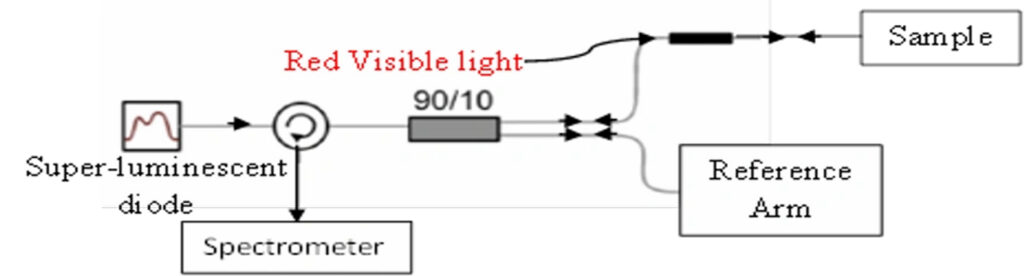
Vibrational Optical Coherence Tomography (VOCT Involves use of near infra-red light and audible sound applied transversely to the skin to produce an image and measure of the resonant frequency of tissue components (Figure 1 as seen above). A diagram of the components that make up the OptoScope are shown in Figure 2 above. The resonant frequency is related to the tissue modulus using a calibration curve developed in the laboratory after testing different tissues.
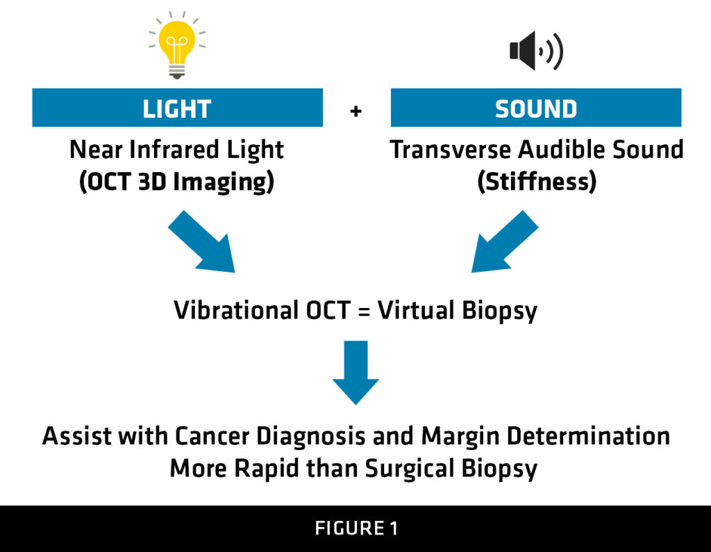
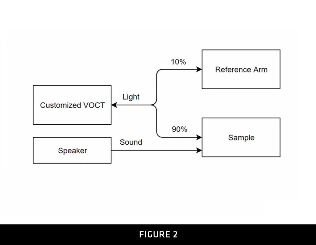
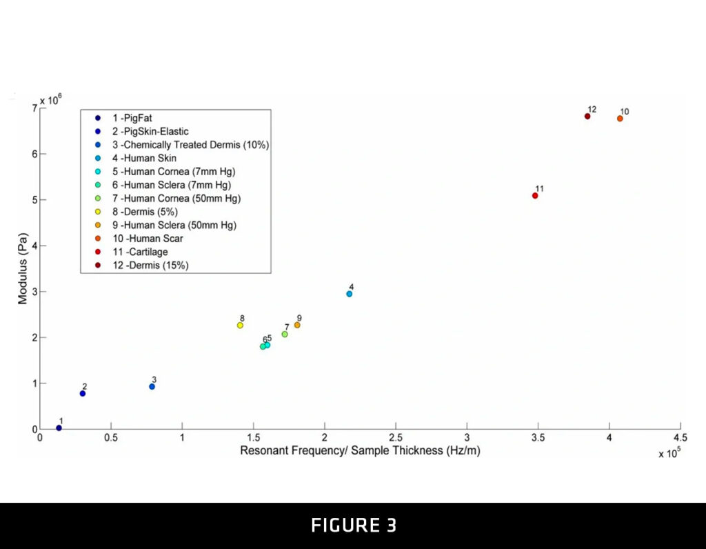
Figure 1. Diagram illustrating the principles of operation of the OptoScope using the principles of vibrational optical coherence tomography (VOCT). Infra-red light from a laser diode, and audible sound from a speaker are applied transversely to a sample surface. With the speaker turned off, the surface of the sample is scanned and an image is recorded. With the OptoScope focused on a single point, the speaker is turned on and the frequency at which the maximum displacement occurs is determined. The modulus of that point is determined from a calibration curve Figure 2 constructed from measurements made using calibration standards and uniaxial tensile tests in vitro as seen in figure 3 above.
Figure 2. Diagram showing the arrangement of components in the OptoScope used to get an OCT image and a measurement of the resonant frequency using audible sound applied transversely to the sample surface. The OptoScope unlike ultrasound and conventional OCT instruments provides an image and physical data at individual points within the image scan. This physical data provides a characteristic “finger print” that can be used to differentiate skin lesions from normal skin and non-invasively assist the surgeon in identifying which lesions need to be excised and how much tissue must be removed.
Figure 3. Vibrational modulus versus resonant frequency per unit sample thickness obtained from vibrational OCT measurements, The resonant frequency is obtained from the frequency at which the maximum deformation is measured after applying audible sound waves at frequencies between 30 and 700 Hz. The thickness is measured from the OCT image. Once the resonant frequency and thickness are measured at a point from VOCT, the modulus can then be calculated from the Y axis of this figure.
ADVANTAGES
- OptoScope 2.0™ is a next-generation material characterization device
- Scans the area of interest in less than 10 minutes from software setup to scan completion
- Sound and light is not felt by patient
- The non-invasive combination of infrared light and transverse vibrational waves create a detailed 2D and 3D image and preserves the area or sample being scanned.
- Measures tissue/material elasticity and viscosity non-invasively. No other characterization technique is known to track material changes in cellular components of human tissue or determine likelihood of failure in non-organic materials.
- OptoScope 2.0™ device can be calibrated against a gold standard

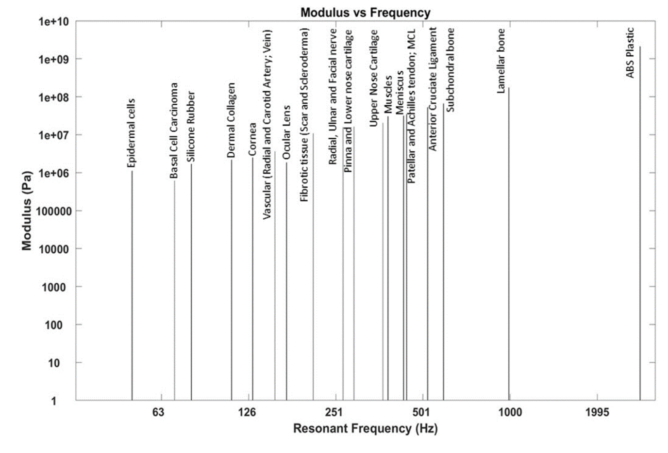
Mechano-Vibrational Spectrum of Surface and Internal Tissues Obtained Using the Vibrational OptoScope
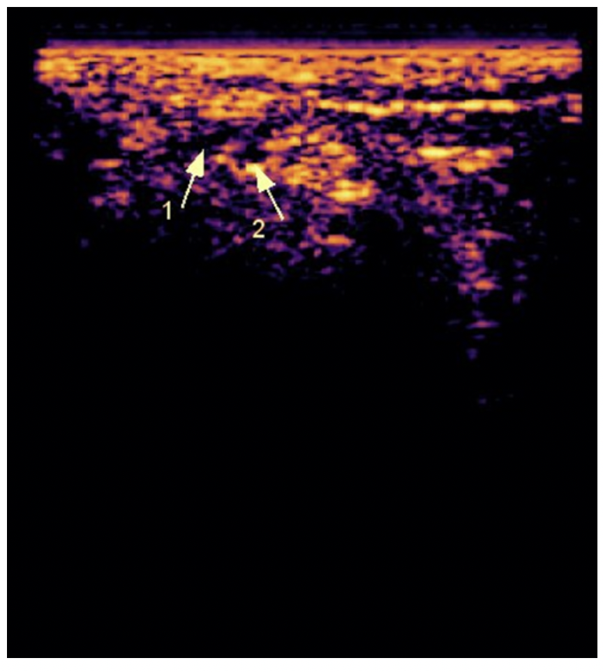
Ultrasound Image of Sural Nerve (1) and Sural Artery (2)